Confocal Microscopy

Laser scanning confocal fluorescence system TCS SP2 AOBS (Leica Microsystems) with inverted microscope DMIRE-2 (Leica)     

Contact: RNDr. Alexandra Zahradníková jr., PhD.

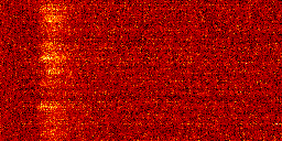
Confocal microscope Leica TCS SP2 is used primarily for measuring local calcium signals during calcium release in the xt mode, when a single line is repeatedly scanned at a rate of up to 2000 lines per second (left image). The recorded image provides data on space (x) and time (t). The confocal microscope is connected to a patch-clamp setup by means of a synchronizer. This arrangement allows controlled stimulation of cells and measurement of the current response and calcium release of the cell in parallel. Fluorescent calcium indicators in combination with non-fluorescent calcium chelators allow imaging of spatially resolved events also when calcium is released in the whole cell volume synchronously.
line-scan image of calcium spikes in rat cardiac myocytes labeling of surface membrane and t-tubules by di-8-ANEPPS
The confocal microscope Leica TCS SP2 can be also used for measuring of static or slow events in the standard xy mode. Fluorescent probes are used to image specific sub-cellular structures such as the surface membrane (right image) and other organelles. Images can be acquired in series of optical slices that allow 3D reconstruction, as well as in time series. Using a right combination of fluorescent indicators, records of both types of measurements (static labeling/dynamic calcium measurement) can be correlated.

Configuration of the confocal microscope Leica TCS SP2:

  • Inverted microscope DM IRE2
  • Objectives 10x and 20x dry, 40x oil, 63x oil, 63x water
  • AOBS, AOTF
  • Scanner 1000 Hz
  • 2x PMT, 1x TLD detectors
  • 1x Ar laser (458, 476, 488, 496, 514 nm)
  • 3x He/Ne laser (543, 594, 633 nm)