
 contact
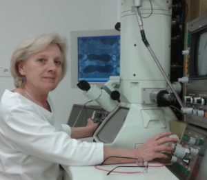
Electron Microscopy
Transmission electron microscope JEM 1200 (Jeol), CCD camera Dual Vision 300W (Gatan)
Contact: RNDr. Marta Novotová, CSc.
Transmission electron microscopy is used for analysis of morphological changes of the cytoarchitecture of cells of animal tissues such as cardiac myocytes, neuronal cells, for studying the architecture of isolated myocytes and cells from tissue cultures (endothelial and glioma cells) as well as to study cellular fractions (myofibrilárna frakcia, reticular and mitochondrial fraction, synaptosomes).
Electron microscope is equipped with a CCD camera and software Gatan Digital Micrograph, what enables acquisition and transfer of image data and enhancement of electron microscopic images, in which the program Adobe Photoshop (Photoshop CS) is also used.
  |
Methods used in electron microscopy
- Fixation of working heart of exprtimental animals on a Langendorff apparatus and processing of the tissue for electron microscopy (isolation of papillary muscle, tissue samples from the left ventricle, fixation, postfication, contrasting, embedding in Durcupan, polymerization).
- Fixation of cells of cell cultures and cellular fractions and their processing for electron microscopy.
- Preparation of ultrathin sections for electron microscopy, contrasting of ultrathin sections.
- Analysis of the ultrastructure of cells of animal tissues (cardiac myocytes, neuronal cells, cultured cells) with the help of the electron microscope. Methodical approach is focused on study of ultrastructural changes at the level of cells and cellular organelles.
- Correlation microscopy that enables to quantify and characterize isolated cardiac myocytes, which have been previously characterized by confocal microscopy, using the electron microscope.
- Image analysis methods for defining changes in volume and surface parameters of microscopic structures and changes in the interaction of cellular organelles. for objective quantification of relationships between cellular organelles we have developed an original stereological method – determination of the neighborhood of organelles. We use our own software Graphic Cell Analyzer (Parulek, Zahradnik, 2009) that uses the principle of the stereological method of vertical sections according to Baddeley (1986).